

Eyes and Vision
Part 1: Eye design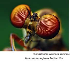
Insects need to deal with the visual world:
This can require using color cues to find food sources
--Vision can be necessary for finding mates, chasing off rivals, and for detecting moving prey
--Visual cues can be essential for finding oviposition sites, and for maintaining circadian rhythms
To perform these functions, the visual organs must receive the information and convey that information to the brain, which then processes the visual world.
 |
 |
|
download video file (.m4v) 28MB |
 Mini-lecture:
The visual system in insects comes in many forms, but the three primary types are:
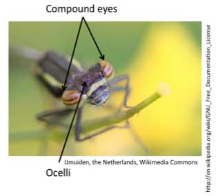
Compound eyes, which are paired structures (normally on either side of the head) composed of many units called ommatidia. Ommatidia are cartridges composed of cuticle, support cells, photoreceptors (cells which detect light), and pigment cells.
Ocelli are single-lens eyes often located on the top of the head (where there are normally three near each other).
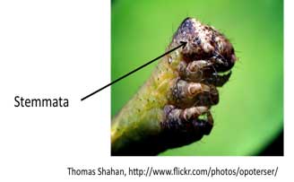
Stemmata are also single-lens eyes which are often found in holometabolous larvae on the sides of the head .
class="greenline">
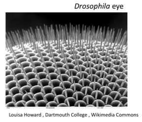
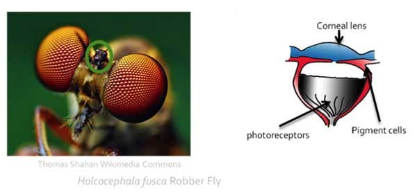
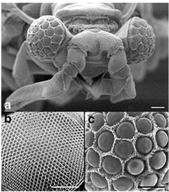
The basic layout: the compound eye
Insects can exhibit a diversity of compound eye types:
From tens of thousands of facets to single facets, to double eyes for living above and below water to single large eyes covering the entire head, insects exhibit massive range of different compound eye designs…
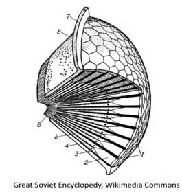
The ommatidia layout: The apposition eye
- The photoreceptors form the core of each ommatidium.
- They can include up to 8 or 9 photoreceptors (also called retinula cells), which are the cells detecting light in the eye.
- The photoreceptors have fine microvilli extending into the middle of the facet, which forms the rhabdom of the eye (which is where the light detecting regions of the eye is located)
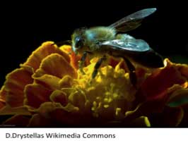
- Many insects have apposition eyes, which include bees and flies
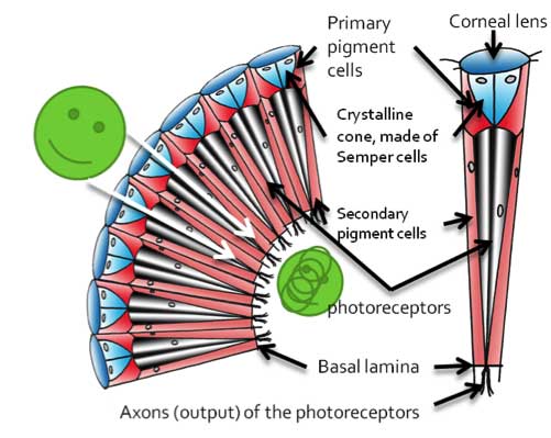
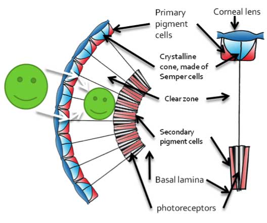
- In this arrangement, photoreceptors form the core of a cartridge of cells which include pigment cells and extend to the crystalline cone.
- Pigment cells surrounding the photoreceptors block light from straying outside of the cartridge of cells, so that each cartridge received light from its own corneal lens.
- Therefore, an image is divided into the different views of the facets of the eye
The ommatidia layout: The superposition eye
- Many insects have superposition eyes, which include beetles, moths, and butterflies
- In this arrangement, photoreceptors are located close to the basal lamina.
- Pigment cells are sequestered to specific areas, allowing light to go through the clear zone to be projected on the rhabdom.
-
Therefore, the image is not divided by the facets of the eye, allowing for more photons to generally reach the photoreceptors.
-
The clear zone can be made up of clear cells or of areas of the photoreceptor cells without pigment (outside of the rhabdom)
-
The superposition eye can gather more light, which is helpful in nocturnal insects such as moths
Photoreceptor layout: different organizational patterns
- The photoreceptors themselves can be arranged in specific ways, where the microvilli (the rhabdom) can be packed together, as is seen in this cross-section of the photoreceptors in the eye (left), called the open rhabdom.
- The microvilli can also be separated (right), which is called the open rhabdom
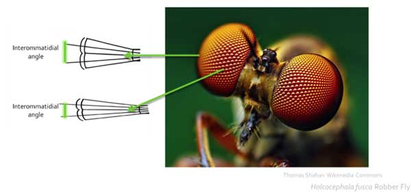
Resolution and the eye: all about structure
- The resolution of the eye depends on the angle between the facets of the compound eye (the interommatidial angle and whether the eye facets are facing the same direction.
- Many insects have high resolution areas, similar to the vertebrate fovea, where the interommatidial angle is low (nearly 1 degree), with the rest of the eye with low resolution (and an increased interommatidial angle)
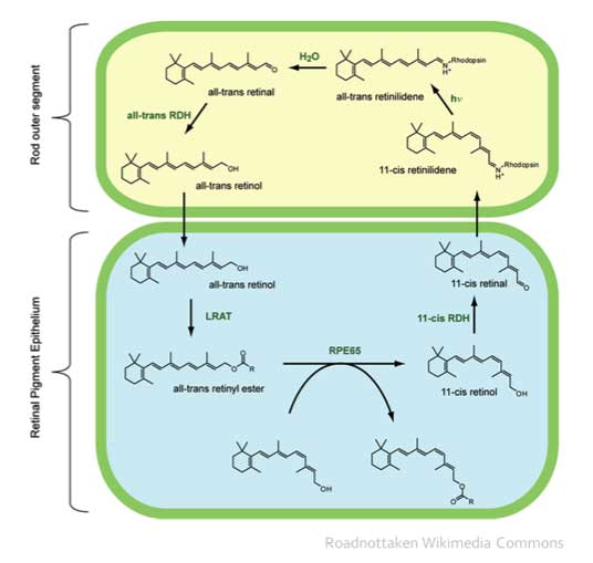
How light is converted into a neural signal
 The phototransduction mechanism is a series of reactions, where a molecule, retinal, changes shape with the introduction of light (in the process below).
This conformational change is then translated into enzymatic changes (see Figure 22.8 in Insects: Structure and Function)
These enzymatic changes induce a G-protein cascade, leading to the opening of channels and depolarization (or excitation) of the photoreceptors
Dark and light adaptation: multiple ways
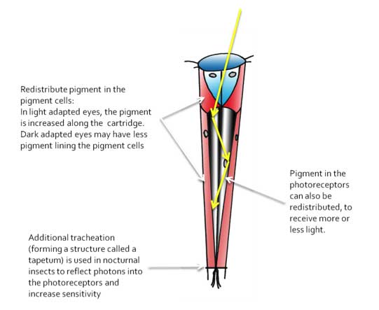
Since insects have to experience a wide range of light conditions, the eye can have a series of adaptations to deal with strong light (called light adaptation) or dim conditions (dark adaptation)
These adaptations are varied, with some adaptations involving mostly nocturnal insects.
In the figure below, photons (yellow arrows) enters the ommatidium, and is reflected off the sides of the cartridge (by the pigment cells), and is hopefully captured and detected by the photoreceptors (center).
The structure of the ocelli
Ocelli are single-lens eyes normally located on the top of the head (where there are normally three near each other)
Since the focus depth for most ocelli indicate they should not form images, ocelli are thought to be primarily light detectors, possibly to detect changes in the horizon.
However, recent recordings in dragonfly ocelli have indicated they can be directionally selective, in an article entitled: Directional Selectivity in the Simple Eye of an Insect, by Joshua van Kleef, Richard Berry, and Gert Stange, in The Journal of Neuroscience, 28(11):2845-2855.
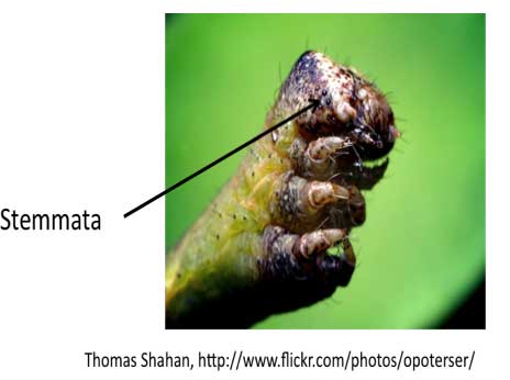
The structure of the stemmata
Stemmata are also single-lens eyes which are often found in holometabolous larvae on the sides of the head
Stemmata are also thought to be poorly developed for image formation, but can be used to detect light.
There are several types of designs, but they tend to be single facets scattered in a group on either side of the head.
Diversity of systems: there’s not just one way to build an eye!
There are a number of other eye modifications, which includes male members of the order Strepsiptera. In these parasites of wasps, the eyes form these eyelets, where images are formed at the level of each eyelet (facet) and are somehow brought together in the brain (see below). Check out the website below by Prof. Elke Buschbeck for other strange modifications of eyes in insects!
 |
|
Elke Buschbeck, University of Cincinnati |
|
 |
TOPIC REVIEWDo you know…?
|