

Integument
Part 2: Glands
|
 Minilecture:
Gland cells
Insects have many different types of glands.
Labial (salivary) glands will be covered in the section on feeding.
Internal glands associated with nervous or other tissues such as malpighian tubules will be covered in sections on hormones and excretion, respectively
A common and diverse set of glands are those associated with the epidermis and are found close to the cuticle and its epidermal cells.
A review of this latter category of epidermal gland cells has classified them into 3 categories: Classes I, II and III (Noirot and Quennedy, 1974).
Class I gland cells are epidermal cells with the outer plasma membrane folded into microvilli. It is assumed that the secretion passes through the cuticle from the microvilli via pore canals. These types of gland cells frequently are clustered in a cuticular region (also called a “gland” but really a conglomeration of gland cells) where their secretions are released as pheromones (see section).
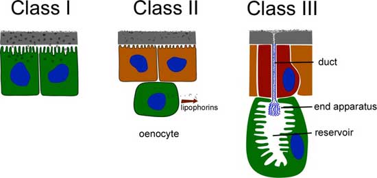 |
Class II gland cells are derived from epidermal cells but don’t have a duct and don’t contact the cuticle. A common type of Class II gland cell are the oenocytes (pronounced ee-no-sites). In flies, they are clustered in the abdomen, just beneath the cuticle and its associated epidermal cells. They release cuticular hydrocarbons which somehow make their way through the cuticle to be released into the waxy layer. It is believed that the secretion binds to lipophorins that carry it through the hemolymph to be released through the cuticle elsewhere. |
J.C.Billener,et al. Nature 461, 987-991 (2009) |
Class III gland cells are the secretory cell of a more complex unit called a class III dermal gland. They are diverse in the function of secretions and their locations on the body, but are always associated with cuticle, hence their name. Dermal glands are multicellular, at least during their development. Sometimes there is only one very large secretory cell present but this is probably due to death (apoptosis) of other cells after their developmental job is done. At least one cell secretes a duct that is lined with a cuticle-like substance. |
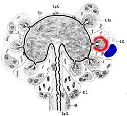
Many class III dermal glands surround the lumen of the colleterial gland of a female sawfly. Image: David Merritt |
Dermal glands & the female reproductive system
Review the structure of the female reproductive system of a generalised insect. The female reproductive tract is ectodermal in origin and cuticle-lined from the external opening to the junction of the ovarian ducts. |
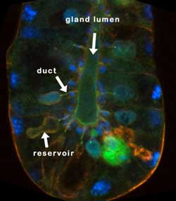
Class III dermal glands surround the lumen of the sperm storage organ (spermatheca) of Drosophila. Image: Modified from Filosi and Pirotti |
Hymenoptera have many dermal glands
Hymenoptera (ants bees and wasps) tend to sociality and consequently they communicate extensively through pheromones. Unsurprisingly they have many sites of pheromone release.
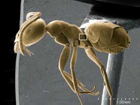
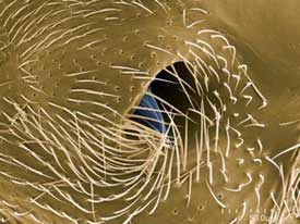
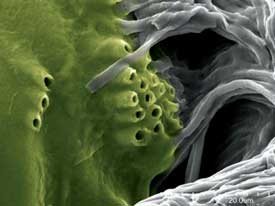
Look in more detail at the metapleural gland of ants found paired on the back of the thorax. One function of its secretion is probably antiseptic as extracts have been shown to kill bacteria, but it has also been suggested that the pheromone communicates communicates an alarm signal to others in the nest. See Roberto Keller’s blog (American Museum of Natural History). The gland ducts open into an atrium that is an in-pocketing of the cuticle. The specimen shown below has hairs within the atrium, although it is not clear whether the secretion is released at the tips or the bases of the hairs.
SEM images: Roberto Keller AMNH
 |
 |
 |
 |
 Go on to the next section to learn about Silks & Secretions
 |
TOPIC REVIEWDo you know…?
|