

Labial Gland Silks 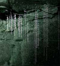
Labial glands are also called salivary glands. Their most common function is to produce saliva, for lubrication of food and initiation of enzyme activity, but in some insects the function has been modified to produce structural materials such as silk, or to produce venoms.
Labial gland silks are the commonest silks. All Lepidoptera produce labial silks. Other lineages have independently evolved labial silks as well. An example is the larvae of the dipteran glow-worm, Arachnocampa. They drop silk lines from a “snare” made of mucous and dot drops of sticky mucous on the silk lines. Flying insects—attracted to the light—are trapped in the silk and mucous and are hauled up and eaten.
In the time-lapse movie below a glow-worm can be seen producing new lines.
Lepidopteran larvae are prodigious silk users. They continuously deposit silk on the substrate such as a plant surface so that if they are dislodged they can climb back up the silken line. They also use silk to form a cocoon around the pupa: the origin of the silk of Bombyx mori, so extensively used by humans.
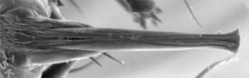
The caterpillar of the agricultural pest moth, Helicoverpa armigera, extrudes silk through a “spigot” below the mandibles. The paired labial glands merge internally before the base of the spigot. The liquid silk precursor protein is forced through a cuticular “silk press” that places the fluid under shearing pressure, causing it to harden into two, connected parallel fibrils.
|
Above right: a typical snare. Below. a glow-worm from SEQ rainforest producing new silk lines, filmed in the laboratory. Time-lapse movie of Arachnocampa flava larva taken at 1 frame every 2 seconds. Images: David Merritt |
|
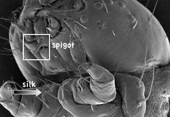
| Above. Spigot of Helicoverpa armigera larva. Bottom. Location of the spigot on the caterpillars head, secreting silk (arrow). Image: Bronwen Cribb |
 |
|
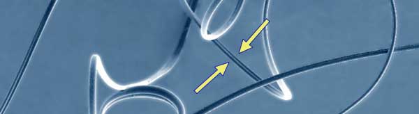
Above. Helicoverpa armigera silk under the SEM, showing it os composed of two, conjoined fibrils. Image: Bronwen Cribb Sorensen GS, Cribb BW, Merritt D, Johnson M-L and Zalucki MP (2006) Structure and ultrastructure of the silk glands and spinneret of Helicoverpa armigera (Hubner) (Lepidoptera: Noctuidae). Arthropod Structure & Development 35:3-13. |
|
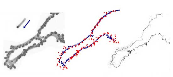
Silk:Above. Movement of a 1st instar Helicoverpa armigera caterpillar and the silk it leaves behind. The three panels show the path of the caterpillar, the position of its head (red dots) and body centre (blue dots) and the silk trail, revealed using a fluorescent stain after the larva was removed. Below is a video of the same event. Images: David Merritt Johnson ML, Merritt DJ, Cribb BW, Trent C and Zalucki MP (2006) Hidden trails: visualizing arthropod silk. Entomologia Experimentalis et Applicata 121:271-274
|